Confocal Microscope
Scientists employ a variety of imaging technologies to visualize and analyze cells on a microscopic level - to reveal what is imperceptible by the naked eye.
These technologies include the classical optical microscope, electron microscopy and laser scanning microscopy. The latter is capable to deliver images of a much higher resolution and contrast than classical optical microscopes. The trick is basically done by having a laser scanning the probe point by point. The reflected light is recorded by a relatively simple device (although of extremely high sensitivity),
a single cell sensor, called photomultiplier (Photomultiplier Tubes = PMT) and then the image is pixel by pixel assembled by a computer / software.
The two-photon laser scanning microscopes even further corroborated the breakthrough in biomedical imaging. The applicability of multi-photon excitation, the optical sectioning capability and the superior contrast of these instruments make them an ideal choice for fluorescence imaging of biological samples.
Gallery: Fluorescence Microscopy
The images in this gallery have been obtained with a confocal LSM (Laser Scanning Microscope) or by conventional fluorescent microscopy. In some cases, the usual "coding" of RGB (red, green, blue) for each channel has been changed to different colors.
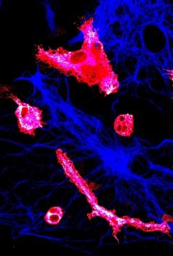
 Astrocytes
GFAP (red), nucleus (blue) tubulin (green)
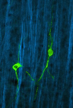
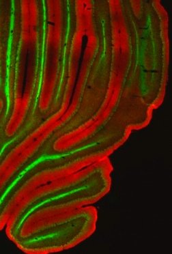 Cerebellum (CNS)
(mouse): neurofilament (green)
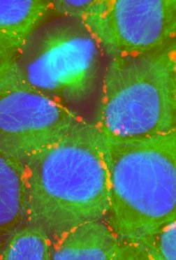
 Hepatocytes (liver cells)
connexin 43 (green), nucleus (red), tubulin (blue)
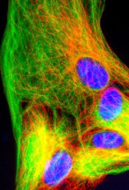
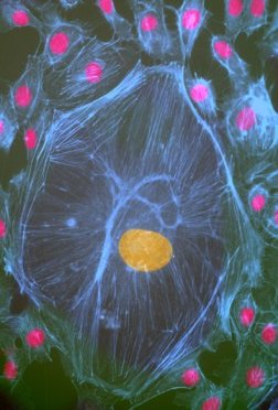
 Hepatocytes (liver cells)
connexin 43 (red), nucleus (blue), tubulin (green); one cell undergoes mitosis
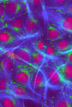
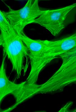 Fibroblasts
nucelus (blue), tubulin (green)
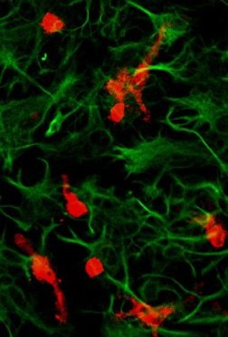 Astrocytes / Microglia
(GFAP, green); microglia (iNOS= inducible nitric oxide synthase (NOS), red)
Move the mouse over the thumbnail, to view an enlarged image in this viewport.
Javascript is not needed, everything has been implemented with css (fluid design: you can use STRG + "scroll wheel" to enlarge the images).
 Hepatocytes (liver cells)
cell nuclei red/yellow, tubulin (blue)
 Astrocytes / Microglia
(blue); microglia (iNOS = inducible nitric oxide synthase (NOS); green), cell nuclei (red)
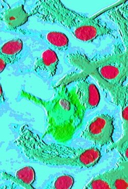
 Microglia
(morphology): Astrocyte (GFAP, bluish), microglia (Ox42, reddish)
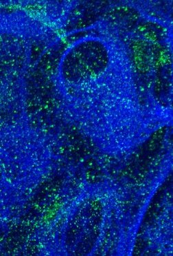 NF-kB in Astrocytes
distribution of NF-kappaB / NF-kB (green) in cell culture (astrocytes (blue))
 NF-kB in Astrocytes
30 minutes after addition of LPS and interferon-gamma: NF-kappaB (NF-kB) is localised mainly within the nucleus
 Cortex (CNS)
(mouse), neurofilament (green), nNOS-positive (neuronal nitric oxide synthase) neurons (red).